
- Greater celandine : Ukrain™, a semisynthetic drug derived from greater celandine (Chelidonium majus), has been studied in clinical trials of various types of cancer with consistently positive outcomes. However, the quality of the research performed to date is inadequate, and higher quality studies are needed.
- Use cautiously in patients taking amphetamines, morphine, hexobarbital, MAOIs, or dopaminergic or serotonergic drugs, or in patients undergoing radiation therapy. Avoid in patients with liver disease or in pregnant and lactating women.
- Guided imagery : Early research suggests that guided imagery may help reduce cancer pain. Further research is needed to confirm these results.
- Guided imagery is usually intended to supplement medical care, not to replace it, and guided imagery should not be relied on as the sole therapy for a medical problem. Contact a qualified health care provider if mental or physical health is unstable or fragile. Never use guided imagery techniques while driving or doing any other activity that requires strict attention. Use cautiously with physical symptoms that can be brought about by stress, anxiety or emotional upset because imagery may trigger these symptoms. If feeling unusually anxious while practicing guided imagery, or with a history of trauma or abuse, speak with a qualified health care provider before practicing guided imagery.
- Meditation : There is good evidence that various types of meditation may help improve quality of life in cancer patients. Studies have shown benefits for mood, sleep quality, and the stresses of treatment. The specific effects of meditation are not fully understood. Additional research is needed in this area.
- Use cautiously with underlying mental illnesses. People with psychiatric disorders should consult with their primary mental healthcare professional(s) before starting a program of meditation, and should explore how meditation may or may not fit in with their current treatment plan. Avoid with risk of seizures. The practice of meditation should not delay the time to diagnosis or treatment with more proven techniques or therapies, and should not be used as the sole approach to illnesses.
- Psychotherapy : Psychotherapy is an interactive process between a person and a qualified mental health care professional (psychiatrist, psychologist, clinical social worker, licensed counselor, or other trained practitioner). There is good evidence that psychotherapy may enhance quality of life in cancer patients by reducing emotional distress and aiding in coping with the stresses and challenges of cancer. Therapy may be supportive-expressive therapy, cognitive therapy or group therapy. While some patients seek psychotherapy in hopes of extending survival, there conclusive evidence of effects on medical prognosis is currently lacking. Psychotherapy may help people come to terms with the fact that they may die of cancer, which is the 4 stage of dealing with a terminal illness, including denial, anger, bargaining, and acceptance.
- Psychotherapy is not always sufficient to resolve mental or emotional conditions. Psychiatric medication is sometimes needed. The reluctance to seek and use appropriate medication may contribute to worsening of symptoms or increased risk for poor outcomes. In order to be successful, psychotherapy requires considerable personal motivation and investment in the process. This includes consistent attendance and attention to treatment recommendations provided by the practitioner. Not all therapists are sufficiently qualified to work with all problems. The client or patient should seek referrals from trusted sources and should also inquire of the practitioner's training and background before committing to work with a particular therapist. Some forms of psychotherapy evoke strong emotional feelings and expression. This can be disturbing for people with serious mental illness or some medical conditions. Psychotherapy may help with post-partum depression, but is not a substitute for medication, which may be needed in severe cases.
- Yoga : Yoga is an ancient system of relaxation, exercise, and healing with origins in Indian philosophy. Several studies report enhanced quality of life in cancer, lower sleep disturbance, decreased stress symptoms and changes in cancer-related immune cells after patients received relaxation, meditation and gentle yoga therapy. Yoga is not recommended as a sole treatment for cancer but may be helpful as an adjunct therapy.
- Yoga is generally considered to be safe in healthy individuals when practiced appropriately. Avoid some inverted poses with disc disease of the spine, fragile or atherosclerotic neck arteries, risk for blood clots, extremely high or low blood pressure, glaucoma, detachment of the retina, ear problems, severe osteoporosis, or cervical spondylitis. Certain yoga breathing techniques should be avoided in people with heart or lung disease. Use cautiously with a history of psychotic disorders. Yoga techniques are believed to be safe during pregnancy and breastfeeding when practiced under the guidance of expert instruction (the popular Lamaze techniques are based on yogic breathing). However, poses that put pressure on the uterus, such as abdominal twists, should be avoided in pregnancy.
- Acupuncture : Acupuncture, or the use of needles to manipulate the "chi" or body energy, originated in China over 5,000 years ago. There has been limited research on acupuncture for cancer pain, and the research that was done was shown to have mixed results. More studies are needed to determine potential benefits. Evidence from several small studies supports use of acupuncture at a specific point on the wrist (P6) to help chemotherapy patients reduce nausea and vomiting. Acupuncture may also reduce the pain associated with cancer.
- Needles must be sterile in order to avoid disease transmission. Avoid with valvular heart disease, infections, bleeding disorders or with drugs that increase the risk of bleeding (anticoagulants), medical conditions of unknown origin, or neurological disorders. Avoid on areas that have received radiation therapy and during pregnancy. Use cautiously with pulmonary disease (like asthma or emphysema). Use cautiously in elderly or medically compromised patients, diabetics or with history of seizures. Avoid electroacupuncture with arrhythmia (irregular heartbeat) or in patients with pacemakers.
- American pawpaw : Evidence supporting the use of the American pawpaw (Asimina triloba) tree for cancer treatment in humans is largely anecdotal and subjective. However, use in humans has reported minimal side effects, and evidence from animal and test tube studies suggest that American pawpaw extract does have some anticancer activity. Pawpaw standardized extract has been used for 18 months in patients with various forms of cancer. Well-designed studies on the long-term effects of pawpaw extracts are currently lacking. Pawpaw should not be used if pregnant or breast-feeding, unless otherwise directed by a doctor.
- Antineoplastons : Antineoplastons are a group of naturally occurring peptide fractions, which were observed by Stanislaw Burzynski, MD, PhD in the late 1970s to be absent in the urine of cancer patients. There is inconclusive scientific evidence regarding the effectiveness of antineoplastons in the treatment of cancer. Several preliminary human studies (case series, phase I/II trials) have examined antineoplaston types A2, A5, A10, AS2-1, and AS2-5 for a variety of cancer types. It remains unclear if antineoplastons are effective, or what doses may be safe. Until better research is available, no clear conclusion can be drawn.
- Avoid if allergic or hypersensitive to antineoplastons. Use cautiously with high medical or psychiatric risk, an active infection due to a possible decrease in white blood cells, high blood pressure, heart conditions, chronic obstructive pulmonary disease, liver disease or damage, or kidney disease or damage. Avoid if pregnant or breastfeeding.
- Arabinoxylan : Arabinoxylan is made by altering the outer shell of rice bran using enzymes from Hyphomycetes mycelia mushroom extract. Arabinoxylan has been found to improve immune reactions in patients with diabetes and cancer of various types. Arabinoxylan products may contain high calcium and phosphorus levels, which may be harmful for patients with compromised renal (kidney) function. Caution is advised when taking arabinoxylan supplements, as numerous adverse effects including drug interactions are possible. Arabinoxylan should not be used if pregnant or breast-feeding, unless otherwise directed by a doctor.
- Aromatherapy : Healing with fragrant oils has been used for thousands of years. Aromatherapy is often used in people with chronic illnesses (frequently in combination with massage), with the intention to improve quality of life or well-being. There is currently not enough scientific evidence to form a firm conclusion about the effectiveness of aromatherapy for quality of life in cancer.
- Essential oils should only be used on the skin in areas without irritation. Essential oils should be administered in a carrier oil to avoid toxicity. Avoid with a history of allergic dermatitis. Use cautiously if driving or operating heavy machinery. Avoid consuming essential oils. Avoid direct contact of undiluted oils with mucous membranes. Use cautiously if pregnant.
- Art therapy : Art therapy involves the application of a variety of art modalities including drawing, painting, clay and sculpture. Art therapy enables the expression of inner thoughts or feelings when verbalization is difficult or not possible. Limited evidence suggests that art therapy may be of benefit in cancer caregiving for families of cancer patients. Possible benefits include reduced stress, lowered anxiety, increased positive emotions and increased positive communication with cancer patients and health care professionals. Art therapy may also reduce pain and other symptoms in cancer patients. More studies are needed to determine how best to use this form of intervention with this population. Art therapy may also benefit children hospitalized with leukemia during and after painful procedures. Limited available study suggests that art therapy improves cooperation with treatment. Children requested art therapy again when procedures were repeated, and parents reported that children were more manageable after art therapy.
- Art therapy may evoke distressing thoughts or feelings. Use under the guidance of a qualified art therapist or other mental health professional. Some forms of art therapy use potentially harmful materials. Only materials known to be safe should be used. Related clean-up materials (like turpentine or mineral spirits) that release potentially toxic fumes should only be used with good ventilation.
- Astragalus : Astragalus (Astragalus membranaceus) has been used in Chinese medicine for centuries for its immune enhancing properties. Although early laboratory and animal studies report immune stimulation and reduced cancer cell growth associated with the use of astragalus, reliable human evidence in these areas is currently lacking. In Chinese medicine, astragalus-containing herbal mixtures are also sometimes used with the intention to reduce side effects of chemotherapy and other cancer treatments. Astragalus-containing herbal combination formulas may also have beneficial effects in aplastic anemia. Due to a lack of well-designed research, a firm conclusion cannot be drawn.
- Caution is advised when taking astragalus supplements, as numerous adverse effects including drug interactions are possible. Astragalus should not be used if pregnant or breast-feeding, unless otherwise directed by a doctor.
- Baikal skullcap : Although the outcomes of early studies using baikal skullcap for cancer are promising, high-quality clinical studies are needed in this area before a conclusion can be made. Avoid if allergic or hypersensitive to Baikal skullcap (Scutellaria barbata), its constituents, or members of the Lamiaceae family. Use cautiously if taking sedatives and/or operating heavy machinery. Use cautiously if taking antineoplastic (anticancer) agents or agents metabolized by cytochrome P450 enzymes. Avoid if pregnant or breastfeeding. Baikal skullcap is an ingredient in PC-SPES, a product that has been recalled from the U.S. market and should not be used.
- Bee pollen : Bee pollen is considered a highly nutritious food because it contains a balance of vitamins, minerals, proteins, carbohydrates, fats, enzymes, and essential amino acids. Research has found that bee pollen may reduce some adverse effects of cancer treatment side effects. Additional study is needed before a firm recommendation can be made. Caution is advised when taking bee pollen supplements as allergic reactions may occur in sensitive individuals. Bee pollen should not be used if pregnant or breast-feeding, unless otherwise directed by a doctor.
- Beta-glucan : Treatment with a beta-glucan, called lentinan, plus chemotherapy (S-1) may help prolong the lives of patients with cancer that has returned or cannot be operated on. More research is needed in this area. Avoid if allergic or hypersensitive to beta-glucan. When taken by mouth, beta-glucan is generally considered safe. Use cautiously with AIDS or AIDS-related complex (ARC). Avoid using particulate beta-glucan. Avoid if pregnant or breastfeeding.
- Bitter melon : Bitter melon (Momordica charantia) is used in Avurvedic medicine from India to lower blood sugar levels. Research has also found that bitter melon extracts may be beneficial in cancer therapies. MAP30, a protein isolated from bitter melon extract, is reported to possess anti-cancer effects in laboratory studies. Potential anti-cancer effects have not been studied appropriately in humans. Caution is advised when taking bitter melon supplements, as numerous adverse effects including blood sugar lowering and drug interactions are possible. Bitter melon should not be used if pregnant or breast-feeding, unless otherwise directed by a doctor.
- Black tea : Black tea (Camellia sinensis) is from the same plant as green tea, but the leaves are processed differently. Black tea usually contains more caffeine than green tea. Several studies have explored a possible association between regular consumption of black tea and rates of cancer in several populations. This research has yielded conflicting results, with some studies suggesting benefits, and others reporting no effects. Laboratory and animal studies report that components of tea, such as polyphenols, have antioxidant properties and effects against tumors. However, effects in humans remain unclear, and these components may be more common in green tea rather than in black tea. Some animal and laboratory research suggests that components of black tea may actually be carcinogenic, or cancer causing, although effects in humans are not clear. Overall, the relationship of black tea consumption and human cancer prevention remains undetermined.
- Avoid if allergic or hypersensitive to caffeine or tannins. Skin rash and hives have been reported with caffeine ingestion. Use caution with diabetes. Use cautiously if pregnant. Heavy caffeine intake during pregnancy may increase the risk of SIDS (sudden infant death syndrome). Very high doses of caffeine have been linked with birth defects. Caffeine is transferred into breast milk. Caffeine ingestion by infants can lead to sleep disturbances/insomnia. Infants nursing from mothers consuming greater than 500 milligrams of caffeine daily have been reported to experience tremors and heart rhythm abnormalities. Tea consumption by infants has been linked to anemia, decreased iron metabolism, and irritability.
- Bovine cartilage : In early study, bovine tracheal cartilage (preparations such as Catrix® and VitaCarte®) has been studied for the treatment of cancer with encouraging results. High quality clinical research is needed to better determine the effectiveness of bovine tracheal cartilage preparations for cancer treatment.
- Avoid if allergic or hypersensitive to bovine cartilage or any of its constituents. Use cautiously with cancer, renal (kidney) failure, or hepatic (liver) failure. Avoid if pregnant or breastfeeding.
- Bromelain : Bromelain is a sulfur-containing digestive enzyme (proteins which help with digestion) that is extracted from the stem and the fruit of the pineapple plant (Ananas comosus). There is not enough information to recommend for or against the use of bromelain in the treatment of cancer, either alone or in addition to other therapies. One small study found that a bromelain supplement decreased tumor size in 12 breast cancer patients. Patients took the supplements for different periods of time, lasting from months to years. Caution is advised when taking bromelain supplements, as numerous adverse effects including blood thinning and drug interactions are possible. Bromelain should not be used if pregnant or breast-feeding, unless otherwise directed by a doctor.
- Cat's claw : Originally found in Peru, the use of cat's claw (Uncaria tomentosa) has been said to date back to the Inca civilization, possibly as far back as 2,000 years. Cat's claw has anti-inflammatory properties, and several low-quality studies suggest that cat's claw may slow tumor growth. However, this research is early and has not identified specific types of cancer that may benefit; thus, the results are not clear. A few studies suggest that cat's claw may also boost the immune system. Caution is advised when taking cat's claw supplements, as numerous adverse effects including blood thinning and drug interactions are possible. Cat's claw should not be used if pregnant or breast-feeding, unless otherwise directed by a doctor.
- Chaparral : Chaparral was used by the Native Americans for various health conditions. The chaparral component nordihydroguaiaretic acid (NDGA) has been evaluated as a treatment for cancer but due to risk of toxicity is considered unsafe and not recommended for use. Chaparral and NDGA have been associated with cases of kidney and liver failure, liver cirrhosis, kidney cysts, and kidney cancer in humans. In response to these reports, the U.S. Food and Drug Administration (FDA) removed chaparral from its "generally recognized as safe" (GRAS) list in 1970. Chaparral and NDGA are generally considered unsafe and are not recommended for use.
- Avoid if allergic to chaparral or any of its components, including nordihydroguaiaretic acid. Use cautiously if taking blood thinners (anticoagulants), blood sugar medication, or drugs that are broken down by the liver (like amiodarone, phenobarbital, valproic acid). Stop use two weeks before surgery/dental/diagnostic procedures with bleeding risk, and do not use immediately after these procedures. Use cautiously if driving or operating heavy machinery. Avoid if pregnant or breastfeeding.
- Chlorella : Chlorella spp. are single-cellular green algae. Clinical study has examined the effect of chlorella on skin cancer. Additional research is needed in this area.
- Avoid in patients with known allergy/hypersensitivity to chlorella, its constituents, or members of the Oocystaceae family. Avoid in patients using warfarin therapy. Avoid long term use or concurrent supplementation with manganese. Use cautiously in patients with hypotension or in patients taking antihypertensives, in patients taking immunomodulators or with altered immune function, in patients with cancer or being treated for cancer, in patients with photosensitivity, taking photosensitizers, or exposed to the sun, or in patients monitoring dioxin or cadmium levels.
- Chlorophyll : Preliminary evidence in suggest that chlorophyll may aid in the reduction of side effects associated with photodynamic therapies, such as those used in management of malignant tumors. Further research is required to support the use of chlorophyll as a laser therapy adjunct for cancer treatment.
- Avoid if allergic or hypersensitive to chlorophyll or any of its metabolites. Use cautiously with photosensitivity, compromised liver function, diabetes or gastrointestinal conditions or obstructions. Use cautiously if taking immunosuppressant agents or antidiabetes agents. Avoid if pregnant or breastfeeding.
- Chrysanthemum : Early study indicates that hua-sheng-ping (includes Chrysanthemum morifolium, Glycyrrhiza uralensis, and Panax notoginseng) may be beneficial for patients with precancerous lesions. However, more research is needed.
- Avoid if allergic or hypersensitive to Chrysanthemum, its constituents, or members of the Asteraceae/Compositae family, such as dandelion, goldenrod, ragweed, sunflower, and daisies. Use cautiously if taking medication for gout, cancer, or HIV. Use cautiously with compromised immune systems or if taking immunomodulators. Avoid with photosensitivity or if taking photosensitizers. Avoid large acute or chronic doses of ingested pyrethrin. Avoid pyrethrin with compromised liver function, epilepsy, or asthma. Avoid ocular exposure to pyrethrin. Avoid if pregnant or breastfeeding.
- Coenzyme Q10Â : Further research is needed to determine if coenzyme Q10 (CoQ10) may be of benefit for cancer when used with other therapies.
- Allergy associated with Coenzyme Q10 supplements has not been reported, although rash and itching have been reported rarely. Stop use two weeks before surgery/dental/diagnostic procedures with bleeding risk and do not use immediately after these procedures. Use caution with a history of blood clots, diabetes, high blood pressure, heart attack, or stroke, or with anticoagulants (blood thinners) or antiplatelet drugs (like aspirin, warfarin, clopidogrel (like Plavix®), or blood pressure, blood sugar, cholesterol or thyroid drugs. Avoid if pregnant or breastfeeding.
- Copper : Copper is a mineral that occurs naturally in many foods, including vegetables, legumes, nuts, grains and fruits, as well as shellfish, avocado, and beef (organs such as liver). Preliminary research reports that lowering copper levels theoretically may arrest the progression of cancer by inhibiting blood vessel growth (angiogenesis). Copper intake has not been identified as a risk factor for the development or progression of cancer. Copper is potentially unsafe when used orally in higher doses than the RDA. Copper supplements should not be used if pregnant or breast-feeding, unless otherwise directed by a doctor.
- Cranberry : Several laboratory studies have reported positive effects of proanthocyanidins, flavonoid components of cranberry (Vaccinium macrocarpon) and other fruits such as blueberries, grape seed, and pomegranate, on health. Based on early laboratory research, cranberry has been proposed for cancer prevention. Additional study is needed in humans before a conclusion can be made.
- Avoid if allergic to cranberries, blueberries or other plants of the Vaccinium species. Sweetened cranberry juice may effect blood sugar levels. Use cautiously with a history of kidney stones. Avoid more than the amount usually found in foods if pregnant or breastfeeding.
- Dandelion : Limited animal research does not provide a clear assessment of the effects of dandelion on tumor growth. Well-conducted human studies are needed to better determine dandelion's effects on cancer.
- Avoid if allergic to chamomile, feverfew, honey, yarrow, or any related plants such as aster, daisies, sunflower, chrysanthemum, mugwort, ragweed, or ragwort. Use cautiously with diabetes or bleeding disorders, gastroesophageal reflux disease (GERD), kidney or liver diseases, or a history of stroke or electrolyte disorders. Monitor potassium blood levels. Stop use two weeks before surgery/dental/diagnostic procedures with bleeding risk and do not use immediately after these procedures. Avoid if pregnant or breastfeeding.
- Echinacea : There is currently a lack of clear human evidence that echinacea affects any type of cancer. The evidence from a small number of clinical trials evaluating efficacy of echinacea in the treatment of radiation-induced leukopenia (decrease in white blood cells) is equivocal. Studies have used the combination product Esberitox®, which includes extracts of echinacea (Echinacea purpurea and pallida) root, white cedar (Thuja occidentalis) leaf, and wild indigo (Baptisia tinctoria) root. Additional clinical studies are needed to make a conclusion.
- Caution is advised when taking echinacea supplements, as numerous adverse effects including drug interactions are possible. Echinacea should not be used if pregnant or breast-feeding, unless otherwise directed by a doctor.
- Essiac® : Essiac® contains a combination of herbs, including burdock root (Arctium lappa), sheep sorrel (Rumex acetosella), slippery elm inner bark (Ulmus fulva), and Turkish rhubarb (Rheum palmatum). The original formula was developed by the Canadian nurse Rene Caisse (1888-1978) and is thought to be effective in cancer therapies, although currently there is not enough evidence to recommend for or against the use of this herbal mixture as a therapy for any type of cancer. Different brands may contain variable ingredients, and the comparative effectiveness of these formulas is not known. None of the individual herbs used in Essiac® has been tested in rigorous human cancer trials, although some components have anti-tumor activity in laboratory studies. Numerous individual patient testimonials and reports from manufacturers are available on the Internet, although these cannot be considered scientifically viable as evidence. Individuals with cancer are advised not to delay treatment with more proven therapies. Caution is advised when taking Essiac® supplements, as numerous adverse effects including drug interactions are possible. Essiac® should not be used if pregnant or breast-feeding, unless otherwise directed by a doctor.
- Focusing : Focusing (experiential therapy) is a method of psychotherapy that involves being aware of one's feelings surrounding a particular issue and understanding the meaning behind words or images conveyed by those feelings. Early evidence suggests focusing may improve mood and attitude in cancer patients. Firm recommendations cannot be made until well-designed clinical trials are available.
- Side effect reporting is rare, but patients should consult with a qualified healthcare practitioner before making decisions about medical conditions and practices. Individuals with severe emotional difficulties should not abandon proven medical and psychological therapies but rather choose focusing as a possible adjunct.
- Folic acid : Folic acid or folate is a form of a water-soluble B vitamin needed for human health. Preliminary evidence suggests that folate may decrease the risk of several types of cancer. Additional research is needed to make a conclusion. Folic acid supplementation may mask the symptoms of pernicious, aplastic, or normocytic anemias caused by vitamin B12 deficiency and may lead to neurological damage.
- Avoid if allergic or hypersensitive to folate or any folate product ingredients. Use cautiously if receiving coronary stents and with anemia and seizure disorders. It is recommended that pregnant women consume 400 micrograms daily in order to reduce the risk of fetal defects. Folate is likely safe if breastfeeding.
- Gamma linolenic acid (GLA)Â : GLA is an omega-6 essential fatty acid. Some laboratory and human studies indicate that GLA may have anti-tumor activity and may be used as a cancer treatment adjunct. Additional research is needed in this area.
- Caution is advised when taking GLA supplements, as numerous adverse effects including an increased risk of bleeding and drug interactions are possible. GLA should not be used if pregnant or breast-feeding, unless otherwise directed by a doctor.
- Garlic : Preliminary human studies suggest that regular consumption of garlic (Allium sativum) supplements may reduce the risk of developing several types of cancer. Some studies use multi-ingredient products so it is difficult to determine if garlic alone may play a beneficial role in cancer prevention. Further well-designed human clinical trials are needed to conclude whether eating garlic or taking garlic supplements may prevent or treat cancer.
- Caution is advised when taking garlic supplements, as numerous adverse effects including an increased risk of bleeding and drug interactions are possible. Garlic should not be used if pregnant or breast-feeding, unless otherwise directed by a doctor.
- Ginseng : Early studies report that ginseng taken by mouth may be of benefit in cancer prevention, especially if ginseng powder or extract is used. Weak studies suggest that ginseng in combination with other herbs may improve cell activity, immune function, and red and white blood cell counts in patients with aplastic anemia; however, other studies have found decreases in blood cell counts. Early studies suggest that ginseng may decrease radiation therapy side effects and may be used as a chemotherapy adjunct to improve body weight, quality of life, and the immune response. There is currently not enough evidence to recommend the use of Panax ginseng or American ginseng for these indications. Study results are unclear, and more research is needed before a clear conclusion can be reached.
- Caution is advised when taking ginseng supplements, as numerous adverse effects including an increased risk of drug interactions are possible. Ginseng should not be used if pregnant or breast-feeding, unless otherwise directed by a doctor.
- Goji : Polysaccharide constituents, such as alpha- and beta-glucans from a variety of plants, are reported to have immune system enhancing properties. In clinical study, Lycium barbarum polysaccharides (LBP) demonstrated a synergistic effect in various cancer treatments, when administered in conjunction with powerful immune stimulating drugs.
- Use cautiously in patients who are taking blood-thinning medications, such as warfarin. Use cautiously in asthma patients and in patients with sulfite sensitivities. The New York Department of Agriculture has detected the presence of undeclared sulfites, a food additive, in two dried goji berry products from China. Avoid in patients who are allergic to goji, any of its constituents, or to members of the Solanaceae family.
- Grape seed : There is currently little information available on the use of grape seed extract in the treatment of human cancer. Further research is needed before a recommendation can be made.
- Avoid if allergic or hypersensitive to grapes or other grape compounds. Use cautiously with bleeding disorders or if taking blood thinners such as warfarin, aspirin, non-steroidal anti-inflammatory drugs (NSAIDS), or anti-platelet agents. Use cautiously with drugs processed using the liver's cytochrome P450 enzyme system. Use cautiously with blood pressure disorders or if taking ACE inhibitors. Avoid if pregnant or breastfeeding.
- Green tea : Green tea is made from the dried leaves of Camellia sinensis, a perennial evergreen shrub. Green tea has a long history of use in health and longevity, dating back to China approximately 5,000 years ago. Although used for centuries to help prevent diseases, the relationship of green tea consumption and human cancer in general remains inconclusive. Evidence from well-designed clinical trials is needed before a firm conclusion can be made in this area.
- Caution is advised when taking green tea supplements, as numerous adverse effects including an increased risk of drug interactions are possible. Green tea should not be used if pregnant or breast-feeding, unless otherwise directed by a doctor.
- Healing touch : Preliminary data suggests that healing touch (HT) may increase quality of life in cancer. However, due to weaknesses in design and the small number of studies, data are insufficient to make definitive recommendations. Studies with stronger designs are needed. HT should not be regarded as a substitute for established medical treatments. Use cautiously if pregnant or breastfeeding.
- Hoxsey formula : "Hoxsey formula" is a misleading name, because it is not a single formula, but rather is a therapeutic regimen consisting of an oral tonic, topical (on the skin) preparations, and supportive therapy. The tonic is individualized for cancer patients based on general condition, location of cancer, and previous history of treatment. An ingredient that usually remains constant for every patient is potassium iodide. Other ingredients are then added and may include licorice, red clover, burdock, stillingia root, berberis root, pokeroot, cascara, Aromatic USP 14, prickly ash bark, and buckthorn bark. A red paste may be used, which tends to be caustic (irritating), and contains antimony trisulfide, zinc chloride, and bloodroot. A topical yellow powder may be used, and contains arsenic sulfide, talc, sulfur, and a "yellow precipitate." A clear solution may also be administered, and contains trichloroacetic acid.
- Well-designed human studies available evaluating the safety or effectiveness of Hoxsey formula are currently lacking. Caution is advised when taking the Hoxsey formula supplements, as numerous adverse effects including an increased risk of drug interactions are possible. Hoxsey formula should not be used if pregnant or breast-feeding, unless otherwise directed by a doctor.
- Hydrazine sulfate : Hydrazine is an industrial chemical marketed as having the potential to repress weight loss and cachexia (muscle wasting) associated with cancer, and to improve general appetite status. However, in large randomized controlled trials, hydrazine has not been proven effective for improving appetite, reducing weight loss, or improving survival in adults. The National Cancer Institute (NCI) sponsored studies of hydrazine sulfate that claimed efficacy in improving survival for some patients with advanced cancer. Trial results found that hydrazine sulfate did not prolong survival for cancer patients. The U.S. Food and Drug Administration (FDA) has received requests from individual physicians for approval to use hydrazine sulfate on a case-by-case "compassionate use" basis on the chance that patients with no other available effective cancer treatment options might benefit from this therapy. The overall controversy in the use of hydrazine sulfate is ongoing, and relevance to clinical practice is unknown. The use of hydrazine sulfate needs to be evaluated further before any recommendations can be made.
- Hydrazine sulfate may cause cancer. Avoid if allergic or hypersensitive to hydrazine sulfate or any of its constituents. Use cautiously with liver or kidney problems, psychosis, diabetes or seizure disorders. Avoid if pregnant or breastfeeding. Side effects have been reported, including dizziness, nausea, and vomiting.
- Iodine : Iodine is an element (atomic number 53), which is required by humans for the synthesis of thyroid hormones (triiodothyronine/T3 and thyroxine/T4). The potential role of non-radioactive iodine in cancer care remains unknown. Antioxidant and anti-tumor effects have been proposed based on laboratory research. In contrast, some scientists have asserted that tumors may uptake more iodine than normal tissues. It has been suggested that high rates of gastric (stomach) cancer or low rates of breast cancer in coastal Japan may be due to high iodine intake, although this has not been demonstrated scientifically. Povidone-iodine solutions have been used as a part of alternative cancer regimens, such as the Hoxsey formula. Preliminary study has also indicated povidone-iodone solution as a potential rectal washout for rectal cancer. Overall, no clear conclusion can be drawn based on the currently available evidence.
- Reactions can be severe, and deaths have occurred with exposure to iodine. Avoid iodine-based products if allergic or hypersensitive to iodine. Do no use for more than 14 days. Avoid Lugol solution and saturated solution of potassium iodide (SSKI, PIMA) with hyperkalemia (high amounts of potassium in the blood), pulmonary edema (fluid in the lungs), bronchitis, or tuberculosis. Use cautiously when applying to the skin because it may irritate/burn tissues. Use sodium iodide cautiously with kidney failure. Avoid sodium iodide with gastrointestinal obstruction. Iodine is safe in recommended doses for pregnant or breastfeeding women. Avoid povidone-iodine for perianal preparation during delivery or postpartum antisepsis.
- Jiaogulan : Preliminary evidence indicates that gypenosides extracted from Gynostemma pentaphyllum may decrease cancer cell viability, arrest the cell cycle, and induce apoptosis (cell death) in human cancer cells. Immune function in cancer patients has also been studied. Additional study is needed in this area.
- Avoid if allergic or hypersensitive to jiaogulan (Gynostemma pentaphyllum), its constituents, or members of the Cucurbitaceae family. Use cautiously with blood disorders or taking anticoagulants or anti-platelet drugs (blood thinners). Use cautiously with diabetes. Avoid if pregnant or breastfeeding.
- Lavender : Perillyl alcohol (POH), derived from lavender (Lavendula officinalis), may be beneficial in the treatment of some types of cancer. Preliminary small studies in humans, involving the use of POH suggest safety and tolerability, but effectiveness has not been established.
- Avoid if allergic or hypersensitive to lavender. Avoid with a history of seizures, bleeding disorders, eating disorders (anorexia, bulimia), or anemia (low levels of iron). Avoid if pregnant or breastfeeding.
- Lutein : Currently, there is insufficient available evidence to recommend for or against the use of lutein for cancer. Available evidence in humans is conflicting.
- Avoid if allergic or hypersensitive to lutein or zeaxanthin. Use cautiously if at risk for cardiovascular disease or cancer. Avoid if pregnant or breastfeeding.
- Lycopene : High levels of lycopene are found in tomatoes and in tomato-based products. Tomatoes are also sources of other nutrients such as vitamin C, folate, and potassium. Several laboratory and human studies examining tomato-based products and blood lycopene levels suggest that lycopene may be associated with a lower risk of developing cancer and may help stimulate the immune system. However, due to a lack of well-designed human research using lycopene supplements, its effectiveness for cancer prevention remains unclear.
- Avoid if allergic to tomatoes or to lycopene. Due to a lack of conclusive data, avoid if pregnant or breastfeeding.
- Maitake mushroom : Maitake is the Japanese name for the edible mushroom Grifola frondosa. Maitake has been used traditionally both as a food and for medicinal purposes. Early studies in the laboratory as well as in humans suggest that beta-glucan extracts from maitake may increase the body's ability to fight cancer. However, these studies have not been well designed, and better research is needed before the use of maitake for cancer can be recommended.
- Caution is advised when taking maitake supplements, as numerous adverse effects including an increased risk of bleeding and drug interactions are possible. Maitake should not be used if pregnant or breast-feeding, unless otherwise directed by a doctor.
- Meditation : Not enough research has shown meditation to be of benefit in cancer prevention. More studies are needed.
- Use cautiously with underlying mental illnesses. People with psychiatric disorders should consult with their primary mental healthcare professional(s) before starting a program of meditation, and should explore how meditation may or may not fit in with their current treatment plan. Avoid with risk of seizures. The practice of meditation should not delay the time to diagnosis or treatment with more proven techniques or therapies, and should not be used as the sole approach to illnesses.
- Melatonin : There are several early-phase and controlled human trials of melatonin in patients with various advanced stage malignancies. There is currently not enough definitive scientific evidence to discern if melatonin is beneficial as a cancer treatment, whether it increases (or decreases) the effectiveness of other cancer therapies, or if it safely reduces chemotherapy side effects.
- Melatonin is not to be used for extended periods of time. Caution is advised when taking melatonin supplements, as numerous adverse effects including drug interactions are possible. Melatonin is not recommended during pregnancy or breastfeeding unless otherwise advised by a doctor.
- Milk thistle : Milk thistle (Silybum marianum) has been used medicinally in China for over 2,000 years, most commonly for the treatment of liver and gallbladder disorders. There are early reports from laboratory experiments that the components silymarin and silibinin found in milk thistle may reduce the growth of human cancer cells. However, effects have not been shown in high-quality human trials.
- Caution is advised when taking milk thistle supplements, as numerous adverse effects including an increased risk of bleeding and drug interactions are possible. Milk thistle should not be used if pregnant or breast-feeding, unless otherwise directed by a doctor.
- Mistletoe : Mistletoe is one of the most widely used unconventional cancer treatments in Europe. Mistletoe extracts have been studied for a variety of human cancers as well as melanoma and leukemia. However, efficacy has not been conclusively proven for any one condition. In fact, some studies have shown lack of efficacy of certain preparations for a variety of cancers. Larger, well-designed clinical trials are needed.
- Caution is advised when taking mistletoe supplements, as numerous adverse effects including nausea, vomiting, and drug interactions are possible. Mistletoe should not be used if pregnant or breast-feeding, unless otherwise directed by a doctor.
- Moxibustion : Moxibustion is a healing technique employed across the diverse traditions of acupuncture and oriental medicine for over 2,000 years. Moxibustion uses the principle of heat to stimulate circulation and break up congestion or stagnation of blood and chi. Moxibustion is closely related to acupuncture as it is applied to specific acupuncture points. Preliminary evidence suggests that moxibustion may reduce side effects of chemotherapy or radiation therapy. More studies are needed.
- Use cautiously over large blood vessels and thin or weak skin. Avoid with aneurysms, any kind of "heat syndrome," cardiac disease, convulsions or cramps, diabetic neuropathy, extreme fatigue and/or anemia, fever, inflammatory conditions, over allergic skin conditions or ulcerated sores, or skin adhesions. Avoid if pregnant or breastfeeding. Avoid areas with an inflamed organ, contraindicated acupuncture points, face, genitals, head, inflamed areas in general, nipples, and skin adhesions. Avoid in patients who have just finished exercising or taking a hot bath or shower. Use cautiously with elderly people with large vessels. It is considered not advisable to bathe or shower for up to 24 hours after a moxibustion treatment.
- Oleander : Laboratory studies of oleander (Nerium oleander) suggest possible anti-cancer effects, although reliable research in humans is not currently available. There are reports that long-term use of oleander may have positive effects in patients several types of cancer. More research is needed.
- Caution is advised when taking oleander supplements, as numerous adverse effects including drug interactions are possible. Oleander should not be used if pregnant or breast-feeding, unless otherwise directed by a doctor.
- Omega-3 fatty acids : Omega-3 fatty acids are essential fatty acids found in some plants and fish. A balance of omega-6 and omega-3 fatty acids is advised for health. Several population studies report that dietary omega-3 fatty acids or fish oil may reduce the risk of developing several different types of cancer. Well conducted clinical trials are necessary before a clear conclusion can be drawn regarding the use of omega-3 fatty acids for cancer prevention.
- Caution is advised when taking omega-3 fatty acid supplements, as numerous adverse effects including an increase in bleeding and drug interactions are possible. Omega-3 fatty acid supplements should not be used if pregnant or breast-feeding, unless otherwise directed by a doctor.
- Para-aminobenzoic acid : N-butyl-p-aminobenzoate (BAB) has been shown to be a lipid-soluble local anesthetic. Early study found significant pain relief in patients with intractable cancer pain after an epidural injection of BAB suspension. Larger scale clinical study is needed to confirm these findings.
- Avoid with known hypersensitivity to PABA or its derivatives. Avoid oral use in children and pregnant or nursing women. Use cautiously in patients with renal disease, bleeding disorders or taking anticoagulants, diabetics or patients at risk for hypoglycemia. Discontinue use if rash, nausea, or anorexia occurs. Pharmaceutical doses of PABA and its derivatives should only be taken under appropriate medical supervision. PABA should not be given concurrently with sulfonamides.
- Perillyl alcohol : Perillyl alcohol has been used to treat cancer. However, high quality scientific studies are lacking. Further research is required before recommendations can be made.
- Avoid if allergic/hypersensitive to perillyl alcohol. Avoid use in the absence of medical supervision. Use cautiously in patients under medical supervision. Avoid if pregnant or breastfeeding.
- Prayer : Initial studies of prayer in patients with cancer (such as leukemia) report variable effects on disease progression or death rates when intercessory prayer is used. Better quality research is necessary before a firm conclusion can be drawn.
- Prayer is not recommended as the sole treatment approach for potentially serious medical conditions, and should not delay the time it takes to consult with a healthcare professional or receive established therapies. Sometimes religious beliefs come into conflict with standard medical approaches, and require an open dialog between patients and caregivers. In clinical study, patients certain that they were receiving intercessory prayer had a higher incidence of complications following cardiac bypass surgery than those who did not know they were being prayed for.
- Reiki : Reiki may contribute to reduced perception of pain, improved quality of life, and reduced fatigue in cancer patients. More studies are needed.
- Reiki is not recommended as the sole treatment approach for potentially serious medical conditions, and should not delay the time it takes to consult with a healthcare professional or receive established therapies. Use cautiously with psychiatric illnesses.
- Reishi mushroom : Reishi (Ganoderma lucidum) has been shown to have antineoplastic and immunomodulatory effects in animal studies. Human studies exist of advanced cancer patients using Ganopoly®, a Ganoderma lucidum polysaccharide extract. Results show improved quality of life and enhanced immune responses, which are typically reduced or damaged in cancer patients receiving chemotherapy and/or radiation therapy. Well-designed long-term studies are needed confirm these results and to determine potential side effects.
- Caution is advised when taking reishi supplements, as numerous adverse effects including an increased risk of bleeding and drug interactions are possible. Reishi should not be used if pregnant or breast-feeding, unless otherwise directed by a doctor.
- Resveratrol : The effects of resveratrol cannot be adequately assessed from trials using foods, wine, or combination products containing resveratrol and other substances. Well-designed clinical trials of resveratrol alone are needed before a recommendation can be made in regards to cancer prevention and/or treatment.
- Avoid if allergic or hypersensitive to resveratrol, grapes, red wine or polyphenols. Resveratrol is generally considered safe and is commonly found in food and beverages. Use cautiously with bleeding disorders, abnormal blood pressure. Use cautiously with drugs that are broken down by the body's cytochrome P450 system or digoxin (or digoxin-like drugs). Avoid if pregnant or breastfeeding.
- Seaweed : Bladderwrack (Fucus vesiculosus) is a brown seaweed that grows on the northern coasts of the Atlantic and Pacific oceans, and the North and Baltic seas. Bladderwrack appears to suppress the growth of various cancer cells in animal and laboratory studies. However, reliable human studies to support a recommendation for use in cancer are currently lacking.
- Caution is advised when taking bladderwrack supplements, as numerous adverse effects including an increased risk of drug interactions are possible. Bladderwrack should not be used if pregnant or breast-feeding, unless otherwise directed by a doctor.
- Selenium : Selenium is a trace mineral found in soil, water, and some foods. It is an essential element in several metabolic pathways. Several studies suggest that low levels of selenium (measured in the blood or in tissues such as toenail clippings), may be a risk factor for developing cancer. Population studies suggest that people with cancer are more likely to have low selenium levels than healthy matched individuals, but in most cases it is not clear if the low selenium levels are a cause or merely a consequence of disease. It currently remains unclear if selenium is beneficial for cancer prevention or cancer treatment.
- Avoid if allergic or sensitive to products containing selenium. Avoid with a history of nonmelanoma skin cancer. Selenium is generally regarded as safe for pregnant or breastfeeding women. However, animal research reports that large doses of selenium may lead to birth defects.
- Shark cartilage : For several decades, shark cartilage has been proposed as a cancer treatment. Studies have shown shark cartilage or the shark cartilage product AE-941 (Neovastat®) to block the growth of new blood vessels, a process called "anti-angiogenesis," which is believed to play a role in controlling growth of some tumors. There have also been several reports of successful treatments of end-stage cancer patients with shark cartilage, but these have not been well-designed and have not included reliable comparisons to accepted treatments. Many studies have been supported by shark cartilage product manufacturers, which may influence the results. In the United States, shark cartilage products cannot claim to cure cancer, and the U.S. Food and Drug Administration (FDA) has sent warning letters to companies not to promote products in this way. Without further evidence from well-designed human trials, it remains unclear if shark cartilage is of any benefit in cancer and patients are advised to check with their doctor and pharmacist before taking shark cartilage.
- Shark cartilage available in Asian grocery stores and restaurants should not be eaten due to declining populations of sharks. Caution is advised when taking shark cartilage supplements, as numerous adverse effects including an increased risk of drug interactions are possible. Shark cartilage should not be used by patients who are pregnant or breast-feeding, unless otherwise directed by a doctor.
- Shiitake mushroom : Shiitake (Lentinus edodes) has been taken by mouth for boosting the immune system, decreasing cholesterol levels, and for anti-aging. Lentinan, derived from shiitake, has been injected as an adjunct treatment for cancer and HIV infection. Laboratory, animal and human studies of lentinan have shown positive results in cancer patients when used as a chemotherapy adjunct. Further well-designed clinical trials on all types of cancer are required to confirm these results.
- Caution is advised when taking shiitake supplements, as numerous adverse effects including an increased risk of bleeding and drug interactions are possible. Shiitake should not be used if pregnant or breast-feeding, unless otherwise directed by a doctor.
- Slippery elm : Slippery elm is found as a common ingredient in a purported herbal anticancer product called Essiac® and a number of Essiac-like products. These products contain other herbs such as rhubarb, sorrel, and burdock root. Currently, there is not enough evidence to recommend for or against the use of this herbal mixture as a therapy for any type of cancer. Avoid if allergic or hypersensitive to slippery elm. Avoid if pregnant or breastfeeding.
- Sorrel : Early evidence suggests that herbal formulations containing sorrel, such as Essiac®, do not shrink tumor size or increase life expectancy in patients with cancer. However, currently there is a lack of studies evaluating sorrel as the sole treatment for cancer. A conclusion cannot be made without further research.
- Avoid with a known allergy or hypersensitivity to sorrel. Avoid large doses due to reports of toxicity and death, possibly because of the oxalate found in sorrel. Many tinctures contain high levels of alcohol and should be avoided when driving or operating heavy machinery. Sorrel formulations may cause nausea or vomiting when taken with the prescription drugs metronidazole (Flagyl®) or disulfiram (Antabuse®). Avoid if pregnant or breastfeeding.
- Soy : Soy (Glycine max) contains compounds which have been reported to be effective as a cancer treatment. Genistein, an isoflavone found in soy, has been found in laboratory and animal studies to possess anti-cancer effects, such as blocking new blood vessel growth (anti-angiogenesis), acting as a tyrosine kinase inhibitor (a mechanism of many new cancer treatments), or causing cancer cell death (apoptosis). In contrast, genistein has also been reported to increase the growth of pancreas tumor cells in laboratory research. Until reliable human research is available, it remains unclear if dietary soy or soy isoflavone supplements are beneficial, harmful, or neutral in people with various types of cancer.
- Caution is advised when taking soy supplements, as numerous adverse effects including an increased risk of drug interactions are possible. Soy should not be used if pregnant or breast-feeding, unless otherwise directed by a doctor.
- Spiritual healing : Cancer patients, especially those who fear recurrence or are unhappy with their physicians, commonly use prayer and spiritual healing. More research is needed to address the effects of spiritual healing on anxiety, depression, and quality of life in patients with cancer.
- Spiritual healing should not be used as the only treatment approach for medical or psychiatric conditions, and should not delay the time it takes to consider more proven therapies.
- Sweet annie : Certain constituents found in sweet annie show promise for use in cancer when used in combination with standard chemotherapy. However, currently there is not enough scientific evidence in humans to make a strong recommendation for this use.
- Avoid if allergic or hypersensitive to sweet annie (Artemisia annua), its constituents, or members of the Asteraceae/Compositae family such dandelion, goldenrod, ragweed, sunflower, and daisies. Use cautiously in patients who are taking angiogenic agents, or recovering from surgery or other wounds. Use cautiously if taking cardiotoxic or neurotoxic agents or with compromised cardiac or neural function. Use cautiously if taking immunostimulants or quinolines. Avoid if pregnant or breastfeeding.
- Transcutaneous electrical nerve stimulation (TENS)Â : Transcutaneous electrical nerve stimulation (TENS) is a non-invasive technique in which a low-voltage electrical current is delivered through wires from a small power unit to electrodes located on the skin. Although TENS has been used with some success in cancer pain, there is not enough reliable evidence to draw a firm conclusion in this area. TENS is often used in combination with acupuncture.
- Avoid with implantable devices, like defibrillators, pacemakers, intravenous infusion pumps, or hepatic artery infusion pumps. Use cautiously with decreased sensation, like neuropathy, and with seizure disorders. Avoid if pregnant or breastfeeding.
- Thiamin (Vitamin B1)Â : Thiamin deficiency has been observed in some cancer patients, possibly due to increased metabolic needs. It is not clear if lowered levels of thiamin in such patients may actually be beneficial. Currently, it remains unclear if thiamin supplementation plays a role in the management of any particular type(s) of cancer.
- Thiamin is generally considered safe and relatively nontoxic. Avoid if allergic or hypersensitive to thiamin. Rare hypersensitivity/allergic reactions have occurred with thiamin supplementation. Skin irritation, burning, or itching may rarely occur at injection sites. Large doses may cause drowsiness or muscle relaxation. Use cautiously if pregnant or breastfeeding.
- Thymus extract : Preliminary evidence suggests that thymus extract may increase disease-free survival and immunological improvement in several types of cancer. Additional study is needed in this area.
- Avoid if allergic or hypersensitive to thymus extracts. Use bovine thymus extract supplements cautiously due to potential for exposure to the virus that causes "mad cow disease." Avoid with an organ transplant or other forms of allografts or xenografts. Avoid if receiving immunosuppressive therapy, with thymic tumors, myasthenia gravis (neuromuscular disorder), untreated hypothyroidism, or if taking hormonal therapy. Avoid if pregnant or breastfeeding.
- Traditional Chinese medicine (TCM)Â : The ancient Chinese philosophy of Taoism provided the basis for the development of Chinese medical theory. TCM uses over 120 different herbs in cancer treatment, depending on the type of cancer and its cause according to Chinese medical theory. Studies have reported significant benefits include reducing tumors, reducing treatment side effects and improved response to treatment. More studies of stronger design are needed before TCM can be recommended with confidence as an adjunct to cancer treatment, although centuries of traditional use in cancer cannot be discounted.
- Chinese herbs can be potent and may interact with other herbs, foods or drugs. Consult a qualified healthcare professional before taking. There have been reports of manufactured or processed Chinese herbal products being tainted with toxins or heavy metal or not containing the listed ingredients. Herbal products should be purchased from reliable sources. Avoid ma huang, which is the active ingredient in ephedra. Avoid ginseng if pregnant or breastfeeding.
- Turmeric : Turmeric (Curcuma longa) is commonly used for its anti-inflammatory properties. Several early animal and laboratory studies report anti-cancer (colon, skin, breast) properties of curcumin. Many mechanisms have been considered, including antioxidant activity, anti-angiogenesis (prevention of new blood vessel growth), and direct effects on cancer cells. Currently it remains unclear if turmeric or curcumin has a role in preventing or treating human cancer. There are several ongoing studies in this area.
- Caution is advised when taking turmeric supplements, as numerous adverse effects including an increased risk of bleeding and drug interactions are possible. Turmeric should not be used if pregnant or breast-feeding, unless otherwise directed by a doctor.
- Vitamin AÂ : Vitamin A is a fat-soluble vitamin, which is derived from two sources: preformed retinoids and provitamin carotenoids. Retinoids, such as retinal and retinoic acid, are found in animal sources such as livers, kidneys, eggs, and dairy products. Carotenoids like beta-carotene (which has the highest vitamin A activity) are found in plants such as dark or yellow vegetables and carrots. It is not clear if vitamin A or beta-carotene, taken by mouth or used on the skin with sunscreen, is beneficial for skin cancer prevention or the treatment of skin cancers or wrinkles.
- At recommended doses, vitamin A is generally considered non-toxic. Avoid if allergic or hypersensitive to vitamin A. Vitamin A toxicity can occur if taken at high dosages. Use cautiously with liver disease or alcoholism. Smokers who consume alcohol and beta-carotene may be at an increased risk for lung cancer or heart disease. Vitamin A appears safe in pregnant women if taken at recommended doses; however, vitamin A excess, as well as deficiency, has been associated with birth defects. Excessive doses of vitamin A have been associated with central nervous system malformations. Use cautiously if breastfeeding because the benefits or dangers to nursing infants are not clearly established.
- Vitamin C (ascorbic acid)Â : Dietary intake of fruits and vegetables high in vitamin C has been associated with a reduced risk of various types of cancer in population studies (particularly cancers of the mouth, esophagus, stomach, colon, or lung). However, it is not clear that it is specifically the vitamin C in these foods that is beneficial, and vitamin C supplements have not been found to be associated with this protective effect. Experts have recommended increasing dietary consumption of fruits and vegetables high in vitamin C, such as apples, asparagus, berries, broccoli, cabbage, melon (cantaloupe, honeydew, watermelon), cauliflower, citrus fruits (lemons, oranges), fortified breads/grains/cereal, kale, kiwi, potatoes, spinach, and tomatoes. Vitamin C has a long history of adjunctive use in cancer therapy, and although there have not been any definitive studies using intravenous (or oral) vitamin C, there is evidence that it has benefit in some cases. Better-designed studies are needed to better determine the role of vitamin C in cancer prevention and cancer treatment.
- Avoid if allergic or sensitive to vitamin C product ingredients. Vitamin C is generally considered safe in amounts found in foods. Vitamin C supplements are also generally considered safe in most individuals if taken in recommended doses. Large doses (greater than 2 grams) may cause diarrhea and gastrointestinal upset. Avoid high doses of vitamin C with glucose 6-phosphate dehydrogenase deficiency, kidney disorders or stones, cirrhosis (inflammation of the liver), gout, or paroxysmal nocturnal hemoglobinuria (bleeding disorder). Vitamin C intake from food is generally considered safe if pregnant or breastfeeding. It is not clear if vitamin C supplements in doses higher than Dietary Reference Intake recommendations are safe for pregnant or breastfeeding women. Vitamin C is naturally found in breast milk.
- Vitamin DÂ : Limited research suggests that synthetic vitamin D analogs may play a role in the treatment of human cancers. However, it remains unclear if vitamin D deficiency raises cancer risk, or if an increased intake of vitamin D is protective against some cancers. Until additional trials are conducted, it is premature to advise the use of regular vitamin D supplementation for cancer prevention.
- Avoid if allergic or hypersensitive to vitamin D or any of its components. Vitamin D is generally well-tolerated in recommended doses; doses higher than recommended may cause toxic effects. Use cautiously with hyperparathyroidism (overactive thyroid), kidney disease, sarcoidosis, tuberculosis, and histoplasmosis. Vitamin D is safe in pregnant and breastfeeding women when taken in recommended doses.
- Vitamin EÂ : Reliable scientific evidence that vitamin E is effective as a cancer treatment is currently lacking.
- Caution is merited in people undergoing chemotherapy or radiation, because it has been proposed that the use of high-dose antioxidants may actually reduce the anti-cancer effects of these therapies. This remains an area of controversy and studies have produced variable results. Patients interested in using high-dose antioxidants such as vitamin E during chemotherapy or radiation should discuss this decision with their medical oncologist or radiation oncologist. Caution is advised when taking vitamin E supplements, as numerous adverse effects including an increased risk of bleeding and drug interactions are possible. Avoid if allergic or hypersensitive to vitamin E. Avoid with retinitis pigmentosa (loss of peripheral vision). Use cautiously with bleeding disorders or if taking blood thinners. Avoid above the recommended daily level in pregnant women and breastfeeding women.
- Apricot : Available clinical trials on the use of whole apricots for cancer are currently lacking. However, some research has been conducted on "Laetrile™," an alternative cancer drug marketed in Mexico and other countries outside of the U.S. Laetrile™ is derived from amygdalin found in apricot pits and nuts such as bitter almond. There are multiple animal studies and initial human evidence to suggest that Laetrile™ is not beneficial in the treatment of cancer. Based on a phase II trial in 1982, the U.S. National Cancer Institute concluded that Laetrile™ is not an effective chemotherapeutic agent. Nonetheless, many people still travel to use this therapy outside the U.S.
- Multiple cases of cyanide poisoning, including deaths, have been associated with Laetrileâ„¢ therapy. Avoid if allergic to apricot, its constituents or members of the Rosaceae family, especially the Prunoideae subfamily of plants. Avoid eating excessive amounts of apricot kernels (about 7 grams daily, or more than ten kernels daily). Use cautiously with diabetes. Use cautiously when taking supplements containing beta-carotene, iron, niacin, potassium, thiamine or vitamin C. Use cautiously when taking products that may lower blood pressure. Avoid if pregnant or breastfeeding.
- Beta-carotene : While diets high in fruits and vegetables rich in beta-carotene have been shown to potentially reduce certain cancer incidences, results from randomized controlled trials with oral supplements do not support this claim.
- There is some concern that beta-carotene metabolites with pharmacological activity can accumulate and potentially have cancer-causing (carcinogenic) effects. A higher, statistically significant incidence of lung cancer in male smokers who took beta-carotene supplements has been discovered. Beta-carotene/vitamin A supplements may have an adverse effect on the incidence of lung cancer and on the risk of death in smokers and asbestos exposed people or in those who ingest significant amounts of alcohol. In addition, high-dose antioxidants theoretically may interfere with the activity of some chemotherapy drugs or radiation therapy. Therefore, individuals undergoing cancer treatment should speak with their oncologist if they are taking or considering the use of high dose antioxidants. Beta-carotene in the amounts normally found in food does not appear to have this adverse effect. Avoid if sensitive to beta-carotene, vitamin A or any other ingredients in beta-carotene products.
- Bitter almond : "Laetrile" is an alternative cancer drug marketed in Mexico and other countries outside of the United States. Laetrile is derived from amygdalin, found in the pits of fruits and nuts such as the bitter almond. Early evidence suggests that laetrile is not beneficial in the treatment of cancer. In 1982, the U.S. National Cancer Institute concluded that laetrile was not effective for cancer therapy. Nonetheless, many people still travel to use this therapy outside the United States.
- Multiple cases of cyanide poisoning, including deaths, have been associated with laetrile therapy. Avoid if allergic to almonds or other nuts. Use cautiously if driving or operating machinery. Avoid if pregnant or breastfeeding because of the risk of birth defects.
- Hypnotherapy, hypnosis : Hypnosis did not reduce radiotherapy side effects such as anxiety and did not improve quality of life in patients undergoing curative radiotherapy in early high-quality studies.
- Use cautiously with mental illnesses like psychosis/schizophrenia, manic depression, multiple personality disorder or dissociative disorders, or with seizure disorders.
- Iridology : There is currently limited available data supporting iridology as a tool for cancer diagnosis. Additional study is needed.
- Iridology should not be used alone to diagnose disease. Studies of iridology have reported incorrect diagnoses, and thus, potentially severe medical problems may go undiagnosed. In addition, research suggests that iridology may lead to inappropriate treatment. Iridology is therefore not recommended as a sole method of diagnosis or treatment for any condition.
- Selenium : Results from the Nutritional Prevention of Cancer (NPC) trial, conducted among 1,312 Americans over a 13-year period, suggest that selenium supplementation is ineffective for nonmelanoma skin cancer prevention and may actually increase the risk of squamous cell carcinoma and total nonmelanoma skin cancer. Therefore, selenium supplementation should be avoided in individuals at risk or with a history of nonmelanoma skin cancer.
- Avoid if allergic or sensitive to products containing selenium. Avoid with history of nonmelanoma skin cancer. Selenium is generally regarded as safe for pregnant or breastfeeding women. However, animal research reports that large doses of selenium may lead to birth defects.
- Vitamin EÂ : Recent evidence from well-conducted clinical study reports no reduction in the development of cancer with the use of natural-source vitamin E taken daily. Previously, there have been laboratory, population, and other human trials examining whether vitamin E is beneficial in general cancer prevention, including that for prostate, colon, or stomach cancer. Results of these prior studies have been variable. At this time, based on the best available scientific evidence, and recent concerns about the safety of vitamin E supplementation, vitamin E cannot be recommended for cancer prevention.
- Avoid if allergic or hypersensitive to vitamin E. Avoid with retinitis pigmentosa (loss of peripheral vision). Use cautiously with bleeding disorders or if taking blood thinners. Avoid above the recommended daily level in pregnant women and breastfeeding women.
medical conditions
Skin Cancer
Explore the available health information, treatment context, and integrative evidence for Skin Cancer.
Evidence Ledger
Grades describe the evidence for Skin Cancer relative to each listed condition or goal.
Toggle evidence section
Evidence and integrative options for Skin Cancer
Background
- Skin cancer is the abnormal growth of skin cells. It most often develops on skin exposed to the sun, but can also occur on areas that are not ordinarily exposed to sunlight. Skin cancer is generally divided into two stages, local (where the cancer affects only the skin) and metastatic (where cancer has spread beyond the skin).
- More than one million new cases of skin cancer will be diagnosed in the United States this year. One in five Americans will develop some form of skin cancer during their lifetime.
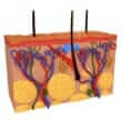
- The skin consists of three layers including the epidermis, dermis, and the subcutaneous layer. The epidermis is the outermost layer and is very thin. It provides a protective layer of skin cells that sheds continually. Squamous cells lie just below the outer surface. Basal cells, which produce new skin cells, are at the bottom of the epidermis. The epidermis also contains cells called melanocytes, which produce melanin, the pigment that gives skin its normal color and causes moles. When exposed to the sun, these cells produce more melanin that helps protect the deeper layers of skin. The extra melanin is what produces the darker color of tanned skin.
- There are three major types of skin cancer, including basal cell carcinoma, squamous cell carcinoma, and melanoma. Basal and squamous cell carcinomas are slow-growing and generally highly treatable. According to the Skin Cancer Foundation, if all forms of skin cancer are found early and treated appropriately, they are all nearly 100% curable. It is very important to limit or avoid exposure to ultraviolet (UV) radiation and pay close attention to suspicious changes in the skin.
- Other less common types of skin cancer include Kaposi's sarcoma, Merkel cell carcinoma, and sebaceous gland carcinoma.
- Basal cell carcinoma: Basal cell carcinoma (BCC) is the most common form of cancer, accounting for nearly 90% of all skin cancers. Basal cells are cells that line the deepest layer of the epidermis. An abnormal growth of cells in this deep layer is known as BCC. Although BCC can usually be diagnosed with a simple biopsy, has a low rate of metastasis, and is fairly easy to treat when detected early, 5-10% of BCCs can be logically aggressive and resistant to treatment. BCC may invade bone and cartilage, and if not treated appropriately and early, it may be very difficult to eliminate.
- Squamous cell carcinoma: Squamous cell carcinoma (SCC) is the second most common form of skin cancer, with over 200,000 new cases per year estimated in the United States. Squamous cells are cells that compose most of the epidermis. An abnormal growth of squamous cells is known as a SCC. Most SCCs are not life-threatening when identified early and treated appropriately, but SCC may become more difficult to treat, can cause disfigurement, and a small percentage may spread (metastasize) to other organs resulting in death.
- Melanoma: Although melanoma is not the most common of the skin cancers, it causes the most deaths. The American Cancer Society estimates that in 2007, there will be 59,940 new cases of melanoma in the United States. Melanoma is a malignant tumor that originates in melanocytes (produce melanin). The majority of melanomas are black or brown, although some melanomas occasionally stop producing pigment and may be skin tone, pink, red, or purple.
Risk Factors and Causes
- Age: Older individuals have a higher risk of developing skin cancer, mainly because many skin cancers develop slowly. Damage by other risk factors that occurred during childhood or adolescence may not become apparent until middle age, but skin cancer can occur in all ages. Basal cell and squamous cell carcinomas are increasing rapidly among women younger than 40.
- Environmental exposure: Exposure to harsh environmental chemicals, including arsenic, cosmetics, and some herbicides increases the risk of skin cancer.
- Fair skin: The less pigment (melanin) the skin contains, the less protection from damaging UV radiation. People with blond or red hair, or light-colored eyes will freckle or sunburn more easily. These individuals are much more likely to develop skin cancer than a person with darker features.
- Family history: Having a parent or a sibling who has developed skin cancer increases the risk of developing skin cancer. Some families are affected by a condition called familial atypical multiple mole melanoma (FAMMM) syndrome. The hallmarks of FAMMM include a history of melanoma in one or more close relatives and having more than 50 moles, some of which are atypical. Because people with this syndrome have an extremely high risk of developing melanoma, frequent screening for signs of skin cancer is crucial.
- Fragile skin: If the skin is burned, injured, or weakened by treatments for other skin conditions, it is more susceptible to sun damage and skin cancer development.
- Moles: Having lots of moles or abnormal moles (called dysplastic nevi) increases the risk of developing skin cancer. The abnormal moles are irregular and generally larger than normal moles, and are more likely than others to become cancerous.
- Personal history: If an individual has developed skin cancer in the past, their risk for developing the disease again is increased. Even basal cell and squamous cell carcinomas that have been successfully removed can recur in the same spot, often within two to three years.
- Precancerous skin lesions: Skin lesions known as actinic keratoses can increase the risk of developing skin cancer. These precancerous skin growths typically appear as rough, scaly patches that range in color from brown to dark pink. Actinic keratoses are most common on the face, lower arms and hands of fair-skinned people whose skin has been sun damaged.
- Sun Exposure: Individuals who spend a considerable amount of time in the sun can develop skin cancer, especially if the skin isn't protected by sunscreen or clothing. Tanning is the skin's injury response to excessive UV radiation, and increases the risk of skin cancer.
- Sunburn: Sunburn is the body's attempt to heal itself from the sun's damaging rays. Every time an individual gets sunburned, there is an increased risk of damaging skin cells and developing skin cancer. One or more severe, blistering sunburns can increase the risk of skin cancer as an adult.
- Sunny or high-altitude climates: Individuals living in sunny, warm climates are exposed to more sunlight than those in colder climates. Higher elevations make UV rays stronger, and may also increase the risk of developing skin cancer.
- Weak immune system: Individuals with weakened immunities are at a greater risk for developing skin cancer. This includes people living with HIV/AIDS, leukemia, and those taking immunosuppressant drugs after an organ transplant.
- Damage in the epidermis to the normal DNA of skin cells may result in new cells growing out of control, and eventually forming an accumulation of cancer cells.
- UV light: The ultraviolet (UV) radiation in sunlight and commercial tanning lamps and beds may cause DNA damage that can lead to skin cancer. UV light is divided into three wavelength bands, including ultraviolet A (UVA), ultraviolet B (UVB), and ultraviolet C (UVC). Only UVA and UVB rays reach the earth. UVC radiation is completely absorbed by the atmospheric ozone. UVA and UVB rays play a role in the formation of skin cancer by causing changes in skin cell DNA, including the development of oncogenes, which are types of genes that can turn a normal cell into a malignant one. UVA penetrates the skin more deeply than UVB.
- Tanning beds deliver high doses of UVA, which makes them especially dangerous. UVA puts an individual at greater risk of skin cancer than spending long hours in the sun because of the deep penetration of the rays.
- Genetic predisposition: Although most cases of skin cancer are related to environmental factors, such as UV exposure, genetics may also play a role. Some people, especially those with fair skin that freckles easily, may be genetically predisposed to developing skin cancer. Therefore, having a family history of skin cancer may increase the risk of developing the disease.
- Other factors: Exposure to toxic chemicals and radiation treatments may also cause skin cancer.
Signs and Symptoms
- Skin cancer develops primarily on areas of sun-exposed skin, including the scalp, face, lips, ears, neck, chest, arms, hands, and on the legs in women. It also can form on areas not receiving as much light such as the palms, spaces between the toes, and the genital area.
- A cancerous skin lesion can appear suddenly or develop slowly, it depends on the type of cancer.
- Basal Cell Carcinoma: Basal cell carcinoma may appear as a flat, scaly red patch, a pearly or waxy bump on the face, ears, or neck (bumps may bleed or develop a crust), a patch with large blood vessels (may look like a birthmark), or a brown or black raised bump.
- Squamous Cell Carcinoma: Squamous cell carcinoma may appear as a flat, scaly red patch (may look similar to a skin rash), a small, smooth, shiny, or waxy bump, a firm, red nodule on the face, lips, ears, neck, hands or arms, or a red or brown, scaly skin patch.
- Malignant Melanoma: Although it can occur anywhere on the body, melanoma appears most often on the upper back or face in both men and women. Malignant melanoma may appear as a new mole, a mole that has gotten larger, a mole that changes color or shape, a mole that bleeds, a mole that itches or causes pain, or a mole with an uneven border or shape. Warning signs of melanoma include a large brownish spot with darker speckles located anywhere on the body, a simple mole located anywhere on the body that changes color, size, feel or that bleeds, or a small lesion with an irregular border and red, white, blue, or blue-black spots on the trunk or limbs. Other signs include shiny, firm, dome-shaped bumps located anywhere on the body and dark lesions on the palms, soles, fingertips and toes, or on mucous membranes lining the mouth, nose, vagina and anus.
- Superficial spreading melanoma: This type of melanoma is the most common type, accounting for about 70% of all cases. Superficial spreading melanoma travels along the top layer of the skin for a fairly long time before penetrating more deeply. It is most common in younger people. The melanoma appears as a flat or slightly raised discolored patch that has irregular borders and is somewhat geometrical in form, with various colors (tan, brown, black, red, blue or white) in it. The melanoma can be found almost anywhere on the body, but is most likely to occur on the trunk in men, the legs in women, and the upper back in both. Other forms of melanoma include lentigo maligna, acral lentiginous melanoma, and nodular melanoma.
- Kaposi's sarcoma: Kaposi's sarcoma is a rare form of skin cancer that develops in the skin's blood vessels and causes red or purple patches on the skin or mucous membranes. Like melanoma, it's a serious form of skin cancer. This form of skin cancer is mainly seen in people with weakened immune systems, such as people with AIDS or taking medications that suppress their immunity (immunosuppressive medications in transplant patients).
- Merkel cell carcinoma: Merkel cell carcinoma is a rare cancer that appears as firm, shiny nodules occurring on or just beneath the skin and in hair follicles. The nodules may be red, pink, or blue and can vary in size from a quarter of an inch to more than two inches. Merkel cell carcinoma is usually found on sun-exposed areas of the head, neck, arms, and legs. Unlike basal and squamous cell carcinomas, merkel cell carcinoma grows rapidly and often spreads to other parts of the body.
- Sebaceous gland carcinoma: Sebaceous gland carcinoma is an uncommon and aggressive form of skin cancer. This form of skin cancer originates in the oil producing glands in the skin. It usually appears as hard, painless nodules that can develop anywhere, but occurs most on the eyelids where they're frequently mistaken for benign conditions.
- Precancerous skin lesions: Precancerous skin lesions, including actinic keratosis, can also develop into squamous cell skin cancer. Actinic keratoses appear as rough, scaly, brown or dark-pink patches. They're most commonly found on the face, ears, lower arms and hands of fair-skinned people whose skin has been damaged by the sun. Other precancerous skin lesions include actinic cheilitis (in the lips), arsenical keratosis (exposure to arsenic), Bowen's Disease (superficial SCC that hasn't spread), and leukoplakia (disease of the mucous membrane).
Diagnosis
- Most doctors recommend a checkup for skin cancer when a new skin growth, a bothersome change in the skin, a change in the appearance or texture of a mole, or a sore that doesn't heal in two weeks appears.
- Biopsy: If the doctor suspects skin cancer, a small sample of the skin (biopsy) will be removed and analyzed. A biopsy can usually be done in a doctor's office using local anesthetic.
- Squamous cell carcinoma: A biopsy is often the only test needed to determine if the individual has squamous cell carcinoma. This cancer doesn't spread, and larger growths may require further testing.
- Basal cell carcinoma: Nodular basal cell carcinoma is the most common type of skin cancer. This tumor usually resembles a smooth, round, waxy pimple, pale yellow or pearl gray, and may vary in size from a few millimeters to one centimeter. The skin covering the nodule is usually so thin that the slightest injury will cause it to bleed. These tumors are often depressed in the middle and display ulcerations. As the tumor grows, it destroys healthy structures in its path, including nerves, muscles, and blood vessels. Large tumors are easily diagnosed, but smaller ones are often difficult to tell from benign skin conditions, such as warts, seborrheic keratoses, moles, psoriasis, or fever sores. Other types of basal cell carcinomas include superficial, sclerosing, pigmented fibroepithelioma, basosquamous carcinoma, and basal cell nevus syndrome.
- Melanoma: Moles, brown spots, and growths on the skin are usually harmless, but may increase the risk of developing melanoma, especially if there are more than 100 moles on the body. Diagnosis of melanoma will need to be made when moles appear to be asymmetric (if a line is drawn through the mole and the two halves don't match), have uneven borders, variety of colors (shades of brown, tan, or black), are large in size, or are changing. The type of melanoma will also be determined.
- Once the type of melanoma has been established, the next step is to classify the disease as to its degree of severity. Melanoma, like other cancers, is classified in stages, and the stage will determine the treatment. Early melanomas (Stages I and II) are localized, and advanced melanomas (Stages III and IV) have spread to other parts of the body, or metastasized. There are also degrees within stages.
- Breslow's thickness: The most important factor in staging a melanoma is the thickness of the tumor, known as Breslow's thickness, and the appearance of microscopic ulcerations (sores). Breslow's thickness measures in millimeters the distance between the upper layer of the epidermis and the deepest point of the tumor's penetration. The thinner the melanoma, the better the chance of a cure. Breslow's thickness diagnoses include: in situ melanoma confined to the epidermis, very thin tumors of less than 1.0 millimeter, thin tumors of 1.01=2.0 mm, intermediate tumors of 2.0-4.0 mm, and thick melanomas of 4.00 mm or more. The presence of microscopic ulcerations moves the tumor into a later stage.
- Clark's level of invasion: Very thin tumors are classified according to Clark's level of invasion, which describes the number of layers of skin penetrated by the tumor. Clark's level I signifies the melanoma occupies only the epidermis. Clark's level II means that the melanoma penetrates to the layer immediately under the epidermis, the papillary dermis, Clark's level III means that the melanoma fills the papillary dermis and may touch the next layer known as reticular dermis, and Clark's level IV is when the melanoma penetrates into the reticular or deep dermis. Clark's level V melanoma invades the subcutaneous fat.
- Stage I: This category is subdivided according to the thickness of the primary (original) tumor. In Stage 1a, the tumor is less than 1.0 mm in Breslow's thickness without ulceration and is in Clark's level II or III. In Stage Ib, the tumor is less than 1.0 mm in Breslow's thickness with ulceration and/or Clark's level III or IV, or it is 1.01 - 2.0 mm in thickness without ulceration.
- Stage II: This category is also subdivided according to gradations in thickness and/or depth and the presence or absence of ulceration. In Stage IIa, the tumor is 1.01 - 2.0 mm in Breslow's thickness with ulceration, or is 2.01-4.0 mm in thickness without ulceration. In Stage IIb, the tumor is 2.01-4.0 mm in Breslow's thickness with ulceration, or is greater than 4.0 mm in thickness without ulceration. In Stage I?c, the tumor is greater than 4.0 mm in Breslow's thickness with ulceration.
- Stage III: When a melanoma is in Stage III or greater, the tumor has either spread to the lymph nodes or to the skin between the primary tumor and the nearby lymph nodes. This can be determined by examining a biopsy of the node nearest the tumor, known as the sentinel node. Such a biopsy is now frequently done when a tumor is more than 1 mm in thickness, or when a thinner melanoma shows evidence of ulceration. In Stage III, the metastasis (spreading) is to the skin or underlying tissue (subcutaneous) for a distance of more than 2 centimeters (1 cm equals 0.4 inch) from the primary tumor, but not beyond the regional lymph nodes. These metastases are microscopic.
- Stage IV: Stage IV melanoma has metastasized to lymph nodes far away from the primary tumor or to internal organs, most often the lung followed in descending order of frequency by the liver, brain, bone, and gastrointestinal tract.
Complications
- Scarring: Some complications of skin cancer can be scars and disfigurement, but it is not usually life-threatening.
- Metastasis: Once a melanoma is diagnosed past Stage III, metastasis may be found increasing the complications for the patient and treatment involved.
- Recurrent skin cancer: Once there has been a diagnosis of skin cancer, a second tumor may be more likely.
Treatment
- Treatment for skin cancer and the precancerous skin lesions known as actinic keratoses varies depending on the size, type, depth and location of the lesions. Often the abnormal cells are surgically removed or destroyed with topical medications. Most treatments require only a local anesthetic and can be done in an outpatient setting. Sometimes no treatment is necessary beyond an initial biopsy that removes the entire growth.
- Freezing: Actinic keratoses and some small, early skin cancers may be destroyed by freezing them with liquid nitrogen (cryosurgery). The dead tissue sloughs off when it thaws. The treatment may leave a small, white scar.
- Excisional surgery: Excisional surgery involves cutting out (excising) the cancerous tissue and a surrounding margin of healthy skin. A wide excision removing extra normal skin around the tumor may be recommended in some cases.
- Laser therapy: A precise, intense beam of light vaporizes growths, generally with little damage to surrounding tissue, minimal bleeding, swelling, and scarring. This procedure may be used to treat superficial skin cancers or precancerous growths on the lips.
- Mohs' surgery: This procedure is used for larger, recurring or difficult-to-treat skin cancers, which may include both basal and squamous cell carcinomas. The skin growth is removed layer by layer, with each examined under a microscope until no abnormal cells remain. This procedure allows cancerous cells to be removed without taking an excessive amount of surrounding healthy skin.
- Curettage and electrodesiccation: This procedure involves the scraping away of cancer cell layers using a circular blade (curet), and the electric needle destroys any remaining cancer cells. This simple, quick procedure is common in treating small or thin basal cell cancers, and generally leaves a small, flat, white scar.
- Radiation therapy: Radiation may be used to destroy basal and squamous cell carcinomas if surgery isn't an option.
- Chemotherapy: Chemotherapy drugs are used to kill cancer cells. For cancers limited to the top layer of skin, creams or lotions containing anti-cancer agents may be applied directly to the skin. Topical drugs can cause severe inflammation and leave scars. Other types of chemotherapy can be used to treat skin cancers that have spread to other parts of the body.
- Imiquimod (IMQ) and/or 5-fluorouracil (5FU) are two topical creams for skin cancers. IMQ works by stimulating the body's immune system to destroy cancerous cells (called topical immunotherapy). 5FU works as a topical chemotherapy, preventing rapidly dividing cells from growing. Both creams can cause redness and inflammation, and need to be used for many weeks to be effective. Occasionally, these creams may be used in addition to surgery for maximal success. Topical therapy allows for a low risk of scarring and may be applied at home.
- Retinoids: Retinoids, such as tretinoin and isotretinoin, are drugs related to vitamin A and are sometimes used off-label to treat or prevent non-melanoma skin cancer. The retinoids may be taken by mouth or applied to the skin, and their use is being studied in clinical trials for treatment of squamous cell carcinoma. Many side effects exist with these medications, including flu-like symptoms and swelling in the feet and ankles (edema).
- Photodynamic therapy: This treatment destroys skin cancer cells with a combination of laser light and drugs that makes cancer cells sensitive to light.
- Biological therapy (also called immunotherapy): Interferon and interleukin-2 are currently under investigation to treat melanoma and non-melanoma skin cancers. These immunotherapy drugs stimulate the immune system to fight the cancer. Other medications applied to the skin, such as imiquimod (Aldara®), enhance the immune system's reaction to the presence of skin cancer.
- Anti-inflammatory medications: The anti-inflammatory drug diclofenac as a topical cream is used off-label as a treatment in actinic keratinosis. Studies report uses for 60 to 90 days with positive results.
Prevention
- Protection from the sun and ultraviolet (UV) rays: Avoid the sun between 10 a.m. and 4 p.m., because the sun's rays are strongest during this period. Try to schedule outdoor activities for other times of the day, even during the winter months or when the sky is cloudy. Sunburns and suntans cause skin damage that can increase the risk of developing skin cancer. Sun exposure accumulated over time may also lead to skin cancer. Five or more sunburns double the risk of developing skin cancer in a lifetime.
- Sunscreen may not filter out all harmful UV radiation, especially the radiation that can lead to melanoma, but they play a major role in an overall sun-protection program. A broad-spectrum sunscreen with a sun protection factor (SPF) of at least 15 is recommended when going outside year-round. For the most protection, apply sunscreen 20 to 30 minutes before sun exposure and reapply it every two hours throughout the day, as well as after swimming or exercising.
- In July 2006, the U.S. Food and Drug Administration (FDA) approved a new over-the-counter sunscreen marketed as Anthelios SX®. The new sunscreen offers better protection from UVA rays than traditional broad-spectrum sunscreens, according to the manufacturer. This may help reduce the risk of various types of skin cancer, including melanoma, basal, and squamous cell carcinomas.
- It's recommended to wear dark, tightly woven clothing that covers the arms and legs, sunglasses (with UVA and UVB protection), and a broad-brimmed hat, which provides more protection than a baseball cap or visor.
- Avoid tanning beds and tan-accelerating agents: Tanning beds emit UVA rays that penetrate deeper into the skin and can cause precancerous skin lesions.
- Sun-sensitizing medications: Some common prescription and over-the-counter drugs, including some antibiotics, some cholesterol, high blood pressure and diabetes medications, birth control pills, nonsteroidal anti-inflammatory drugs (NSAIDS) such as ibuprofen, and the acne and the chemotherapy agent isotretinoin can make the skin more sensitive to sunlight. Also, some herbal supplements may increase sun sensitivity, such as St. John's wort, commonly used for mild depression. A pharmacist can help determine medications that may cause sun sensitivity.
- Regular check-ups: Examining the skin often for new skin growths or changes in existing moles, freckles, bumps and birthmarks is very important in preventing all forms of skin cancer. Healthcare professionals recommend a complete skin exam every year if an individual is older than 40, or more often if they are at high risk of developing skin cancer.
References
Natural Standard developed the above evidence-based information based on a thorough systematic review of the available scientific articles. For comprehensive information about alternative and complementary therapies on the professional level, go to www.naturalstandard.com. Selected references are listed below.
- American Academy of Dermatology. . Accessed April 28, 2009.
- American Academy of Family Physicians. . Accessed April 28, 2009.
- American Cancer Society. . Accessed April 28, 2009.
- Fecher LA, Cummings SD, Keefe MJ, et al. Toward a molecular classification of melanoma. J Clin Oncol. 2007 Apr 20;25(12):1606-20. View Abstract
- Gimotty PA, Elder DE, Fraker DL, et al. Identification of high-risk patients among those diagnosed with thin cutaneous melanomas. J Clin Oncol. 2007 Mar 20;25(9):1129-34. View Abstract
- Markovic SN, Erickson LA, Rao RD, et al. Malignant melanoma in the 21st century, part 2: staging, prognosis, and treatment. Mayo Clin Proc. 2007 Apr;82(4):490-513. View Abstract
- National Cancer Institute (NCI). . Accessed April 28, 2009.
- Natural Standard: The Authority on Integrative Medicine. . Copyright © 2009. Accessed April 28, 2009.
- Satzger I, Schenck F, Thol F, et al. Therapeutic use of erythropoietin in dermatooncology. J Dtsch Dermatol Ges. 2007 Apr;5(4):280-5. View Abstract
- Scope A, Benvenuto-Andrade C, Agero AL, et al. Correlation of dermoscopic structures of melanocytic lesions to reflectance confocal microscopy. Arch Dermatol. 2007 Feb;143(2):176-85. View Abstract
- Skin Cancer Foundation. . Accessed April 28, 2009.